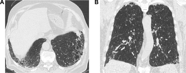
Figure 2.
A 57-year-old former male smoker with clinical chronic obstructive lung disease (COPD).
Notes: (A) Both study observers jointly considered the thick-walled low attenuation areas in the lower lobes as consistent with reticular opacities superimposed to pulmonary emphysema (ie, not as honeycombing). (B) The latter was also abundant in the upper middle lung regions. The LDCT pattern was indeed rated as possible UIP.
Abbreviations: LDCT, low-dose computed tomography; UIP, usual interstitial pneumonia.

