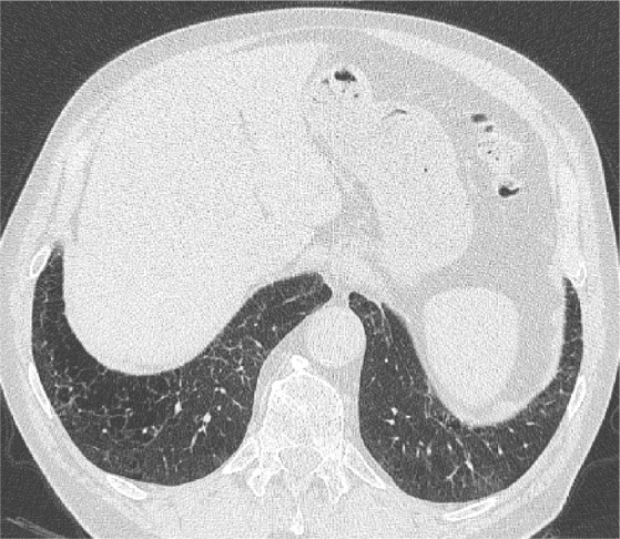
Figure 5.

An axial LDCT image of a 79-year-old former male smoker with clinical COPD showing thick-walled, bizarre-shaped emphysematous areas in the right lower lobe that were regarded as consistent with AEF.
Abbreviations: LDCT, low-dose computed tomography; AEF, airspace enlargement with fibrosis.
