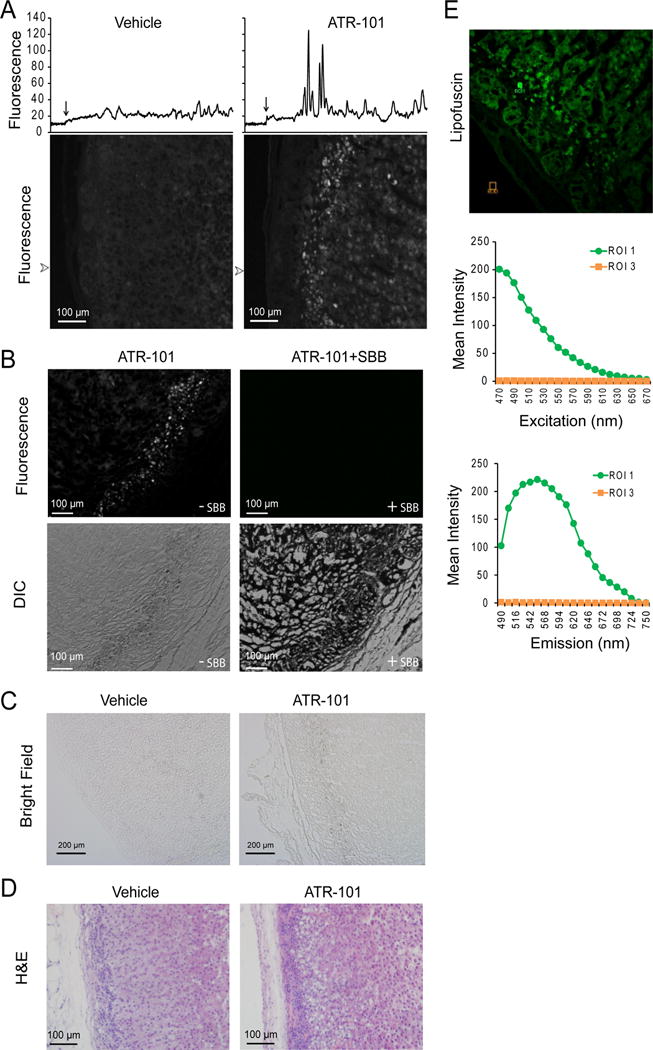
Figure 10. Lipofuscin fluorescence in the zona fasciculata of guinea pigs fed ATR-101.

(A) ATR-101 administration to guinea pigs causes autofluorescence of the zona fasciculata. The panels show autofluorescence images of sections of the adrenal cortex from guinea pigs that were administered vehicle or ATR-101 for two weeks. The traces above the images show the fluorescence intensities along lines traversing from the capsule through the zona reticularis at the arrowheads to the left of the images. The location of the edge of the capsule is shown by an arrow above each trace.
(B) The zona fasciculata layer of guinea pigs administered ATR-101 contains granules and its autofluorescence is quenched by Sudan Black staining. The upper panels show autofluorescence and the lower panels show differential interference contrast images of adjacent sections of the adrenal cortex from a guinea pig that was administered ATR-101 for two weeks. The sections on the right were stained using Sudan Black, and the sections on the left were unstained.
(C) The granules in the zona fasciculata layer of guinea pigs administered ATR-101 are pigmented. The panels show transmitted light images of sections of the adrenal cortex from guinea pigs that were administered vehicle or ATR-101 for two weeks.
(D) The autofluorescent zona fasciculata layer of guinea pigs that were administered ATR-101 overlaps a region of adrenalytic vacuolation. The panels show images of H&E stained sections of the adrenal cortex from guinea pigs that were administered ATR-101 or vehicle for two weeks.
(E) The spectrum of the autofluorescence in adrenals from guinea pigs that were administered ATR-101. The autofluorescence excitation and emission spectra of the regions indicated in the image on the left were analyzed using a Leica SP5X Inverted 2-Photon FLIM Confocal microscope.
The images shown in each panel are representative of adrenacortical sections from 6 guinea pigs that were administered ATR-101 (0.1 g/kg/day po) and from 5 control guinea pigs that were administered vehicle.
