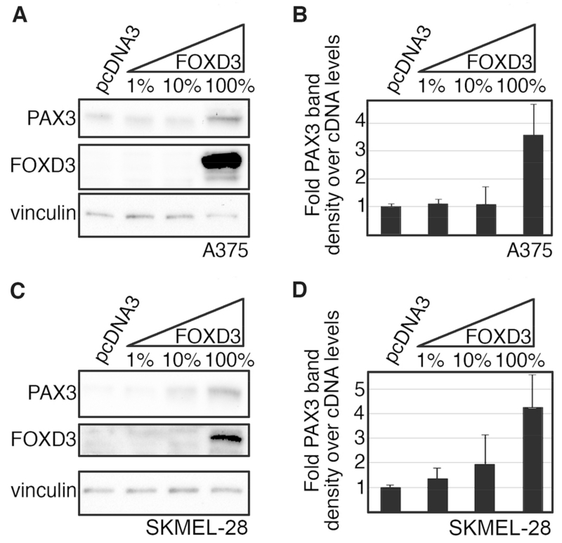
Fig. 2.
FOXD3 is sufficient to drive PAX3 expression in melanoma cells. (A-D) Increasing levels of exogenous FOXD3 leads to an increase in PAX3 in A375 (A,B) and SKMEL-28 (C,D) melanoma cells. Protein levels of PAX3, FOXD3 and vinculin were measured by western analysis in cells transfected with DNA containing increasing levels (1%, 10%, 100%) of FOXD3 expression construct (A,C). Corresponding densitometric analysis for each western blot is shown (B,D). Due to high levels of exogenous expression, endogenous levels of proteins may not be visible in some samples.

