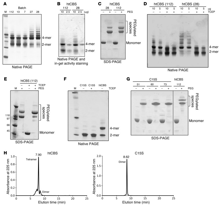
Figure 5. HtCBS C15S mutant prevents protein aggregation, forms mainly dimers, and exhibits a reproducible PEGylation pattern.
(A) Coomassie-stained native PAGE of different htCBS batches. (B) In-gel activity assay for htCBS. (C) Coomassie-stained SDS-PAGE showing a different PEGylation pattern for 2 independent batches of htCBS. (D) Coomassie-stained native PAGE of 2 different htCBS batches, with or without TCEP. (E) Coomassie-stained SDS-PAGE showing the effect of TCEP on PEGylation. (F) Coomassie-stained native-PAGE showing the difference between htCBS C15S and the htCBS dimer/tetramer ratio. (G) Coomassie-stained SDS-PAGE showing the reproducibility of htCBS C15S PEGylation between different batches and comparison with htCBS. (H) SEC-HPLC showing a predominantly tetrameric htCBS and an exclusively dimeric htCBS C15S mutant. M, molecular weight marker.

