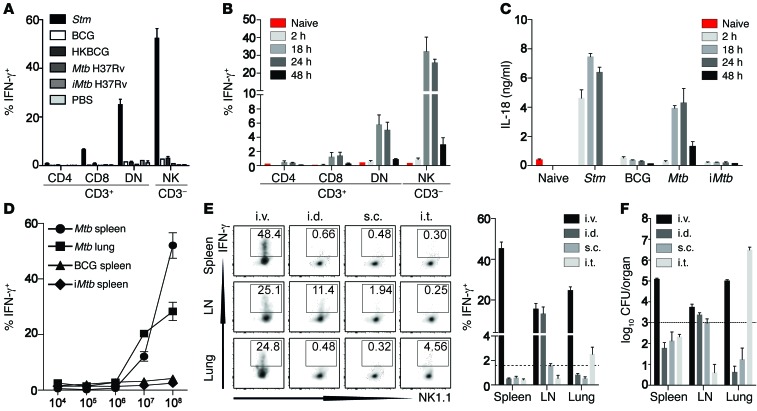
Figure 2. Innate IFN-γ secretion depends on IL-18 and requires Mtb viability.
(A) Percentage of IFN-γ+ cells among total viable splenic CD3+CD8+, CD3+CD4+, CD3+CD4–CD8– (DN) T cells and CD3–NK1.1+ cells 2 hours after B6 mice were injected with 1 × 10⁸ CFU of either Stm (as a positive control), BCG, HKBCG, Mtb H37Rv, iMtb, or PBS. (B) Percentage of IFN-γ+ cells among total viable splenic CD3+CD8+, CD3+CD4+, CD3+CD4–CD8– (DN) T cells and CD3–NK1.1+ cells at different time points after B6 mice were injected with 1 × 10⁸ CFU Mtb H37Rv. (C) Serum IL-18 concentrations at different time points after injection of B6 mice with 1 × 10⁸ CFU Stm, BCG, Mtb H37Rv, or iMtb H37Rv. (D) Percentage of IFN-γ+ cells among total viable CD3–NK1.1+ in spleen and lung 24 hours after injection of different doses of Mtb H37Rv, BCG, or iMtb H37Rv. (E and F) Percentage of IFN-γ+ CD3–NK1.1+ cells (E) and recoverable CFU (F) from either spleen, lung, or draining LN 24 hours after injection of 1 × 10⁸ CFU Mtb H37Rv via the i.v., i.d., i.t., or s.c. route. Results are presented as pooled data (mean ± SEM) (A–F) and representative FACS plots (E) of 5 to 9 (A), 5 to 10 (B and C), 5 (D), or 7 to 10 (E and F) mice per group from at least 2 to 3 pooled, independent experiments. Dotted lines indicate the mean percentage of the smallest reliably detectable IFN-γ+ response by CD3–NK1.1+ cells (E) and the respective mean recoverable CFU (F) 24 hours after Mtb exposure.

