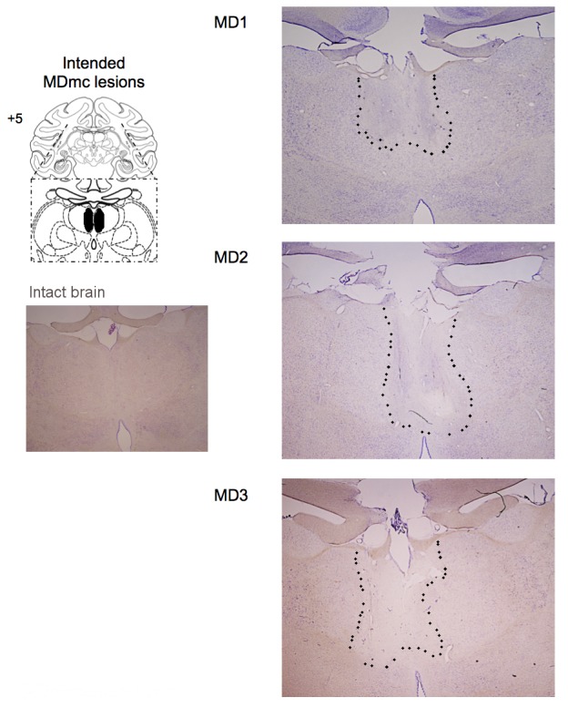
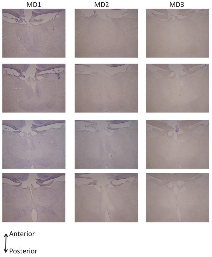
Figure 2. Histological reconstruction of the MDmc lesions.
Coronal sections (right) corresponding to the schematic diagram (left) with lesion detailed (dotted outline) for the bilateral magnocellular mediodorsal thalamic neurotoxic lesions (MDmc) for the three monkeys, MD1, MD2 and MD3. A corresponding coronal section from an intact monkey has been included for comparison.


