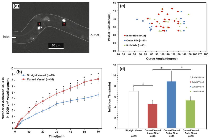
Fig. 2.
a Photomicrograph of MDA-MB-231 cancer cell adhesion to a curved post-capillary venule of diameter ~40 μm after ~30 min perfusion. The bright spots are adherent tumor cells; b comparison of tumor cell adhesion in straight and curved vessels. Data presented are mean ± SE. *p < 0.03; c location of initial tumor cell adhesion in curved vessels as a function of curve angles and vessel diameters; d comparison of initiation times for tumor cell adhesion in straight vessels, at inner, outer and both sides of curved vessels. Data presented are mean ± SE. *p < 0.05; #p < 0.01

