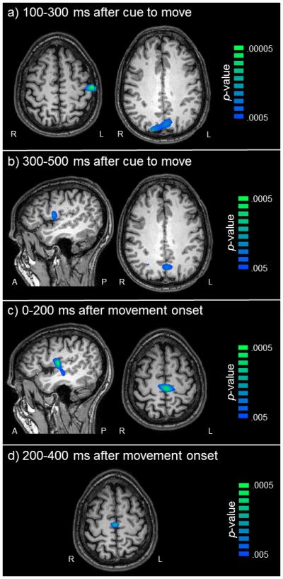
Figure 5. Brain regions with stronger beta ERD during the short planning time condition.

Color bars to the right denote image thresholds (in uncorrected p-values) and all images reflect areas of stronger beta ERD in the short compared to the long condition. Axial slices are shown in radiologic convention (right = left), and all sagittal slices are taken from the right hemisphere. a) Movement Cue 1. Participants exhibited stronger beta ERD in the left postcentral gyrus (left panel) and bilateral parietal regions (right panel) during the short condition from 0.1-0.3 s after the cue to move (p < 0.05, corrected). b) Movement Cue 2. Differences in the right premotor cortex emerged (left panel) and those in the parietal regions persisted from 0.3-0.5 s after the cue to move (right panel; p < 0.05, corrected). c) Movement Onset 1. Significant differences in the right premotor cortex (left panel) were also found during movement onset (p < 0.05, corrected) and marginal differences emerged in the supplementary motor area (SMA) from 0.0-0.2 s after movement onset (SMA; right panel; p = 0.059, corrected; p = .0006, uncorrected). d) Movement onset 2. Marginal differences in beta SMA activity continued 0.2-0.4 s after the onset of movement execution, (p = .07, corrected; p = .0008, uncorrected).
