
Figure 1. GM prevents glutamate-induced oxidative toxicity in HT-22 cells.
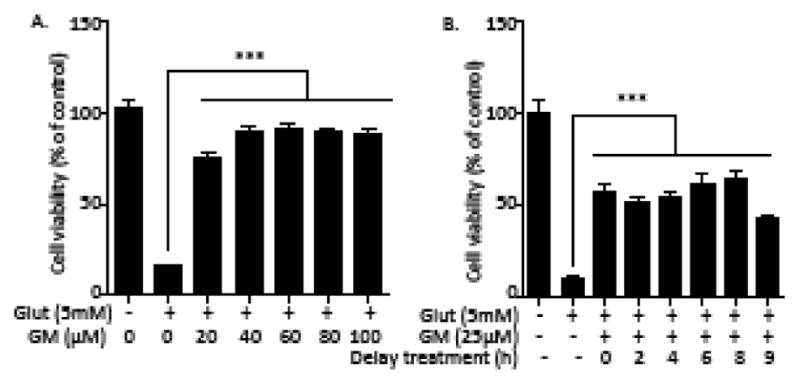
(A) HT-22 cells were exposed to glutamate (5 mM) and GM (20–100 μM) for 24 h. Cell viability was detected by a Calcein AM assay (n=8). (B) Cells were exposed to glutamate (5 mM) for 24 h. GM (25 μM) exposure was applied at 0, 2, 4, 6, 8, 9h after glutamate exposure. Cell viability was detected by aCalcein AM assay (n=8). Representative experiments were independently repeated three times. Results are reported as mean ± SEM. ***P < 0.001 compared with glutamate-treated cells (one-way ANOVA, Tukey’s test).