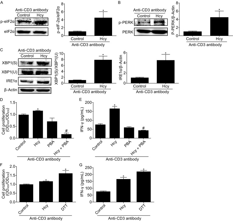
Figure 4.

Hcy enhances T cell ER stress. T cells were incubated with or without Hcy (50 μmol/L) for 4 h with anti-CD3 antibody. Western blot analysis of phosphorylated eIF2α (A), phosphorylated PERK (B), and protein levels of IRE1α and spliced (S) and unspliced (U) XBP1 (C) in T cells with or without Hcy stimulation. β-Actin as a protein loading control. (D) CCK8 assay of cell proliferation with Hcy stimulation with or without ER stress inhibitor PBA or (F) ER stress stimulator DTT. ELISA of IFN-γ with Hcy stimulation with or without PBA (E) or DTT (G). Data are mean ± SEM from 3 independent experiments. *: P < 0.05 vs. Control. #, P < 0.05 vs. Hcy
