
Fig. 1.
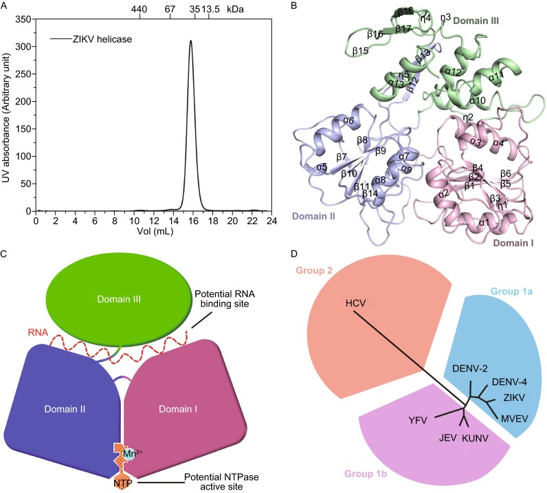
The monomeric structure of ZIKV helicase. (A) Size-exclusion chromatograms of ZIKV helicase. The molecular masses of protein standards are indicated at the top. (B) The overall structure of ZIKV helicase with the three domains colored and labeled respectively. (C) A cartoon diagram illustrating of the overall fold with potential RNA binding site and NTPase active site labelled. (D) Structure-based phylogenetic tree of 8 viral helicase structures from the Flaviviradae family using the program SHP (Stuart et al., 1979) and PHYLIP (Felsenstein, 1997). The following structures with PDB ID in parentheses are included: DENV-2 (2BMF), DENV-4 (2JLQ), JEV (2Z83), KUNV (2QEQ), YFV (1YKS), MVEV (2V8O), HCV (1HEI)