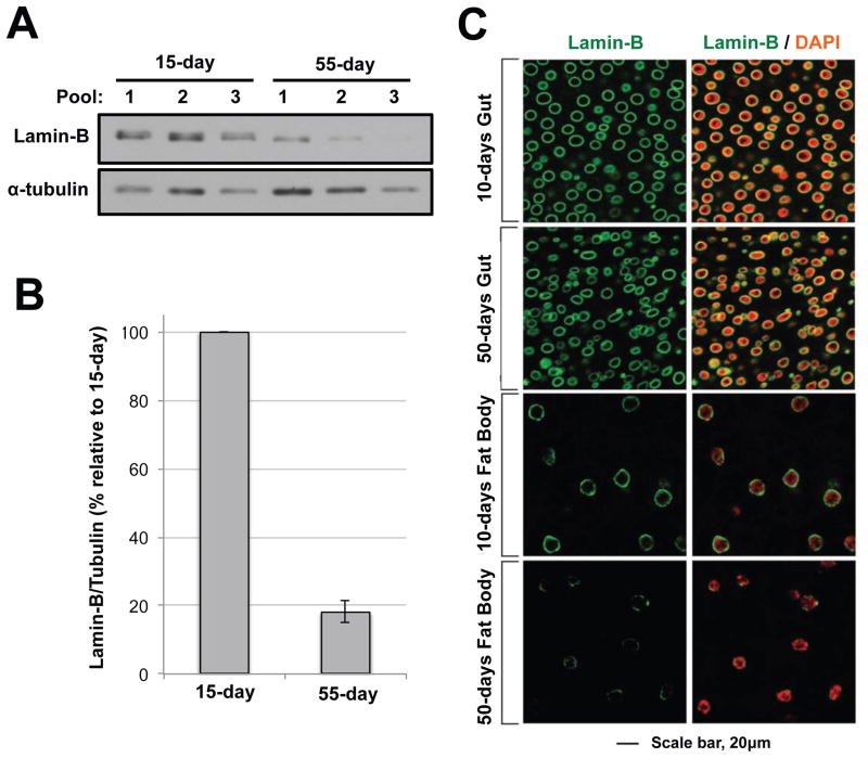
Figure 1. Reduction of lamin-B in the brain and fat body but not in the midgut upon aging.
(a) Lamin-B reduction in old fly brains. Brains were dissected from 15-day or 55-day fruit flies. Five brains were pooled for each sample. Alpha tubulin was used as a loading control. (b) Quantitation of lamin-B western blots. Lamin-B signal was normalized against alpha tubulin, and the levels in the 55-day sample were compared relative to the 15-day sample. (c) Lamin B immunostaining of the midgut (top) and fat body (bottom) from 10 and 50-day old flies. Cells from the 50-day old fat body show reduced and patchy lamin-B staining.

