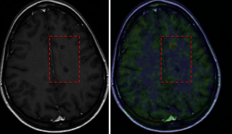
Fig. 1.
In vivo differentiation of chronic T1 lesions using TSPO-PET. Left image a T1-weighted MRI image with two similar-looking (non-gadolinium-enhancing) T1 black holes. TSPO-PET (on the right) shows that in the upper lesion there is microglial activation, confirming this lesion to be a chronic active lesion, whereas in the lower lesion there is no radioligand uptake, confirming this lesion to be a chronic inactive lesion

