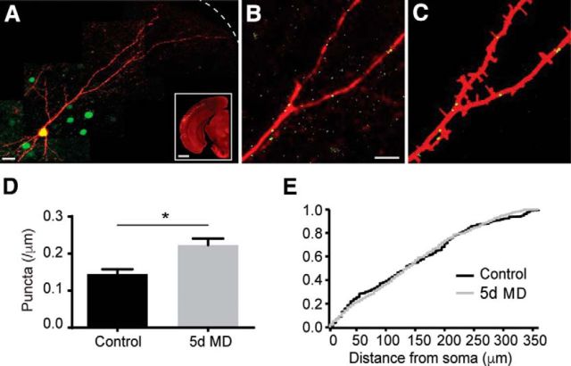
Figure 3.
MD increases gephyrin puncta density in apical dendrites of layer 2/3 PNs. A, dsRED-express (red) and GPHN.FingR staining (green) along the somatodendritic axis of a layer 2/3 PN. Dashed line indicates pial surface. Scale bar, 10 μm. Inset, In utero electroporation of layer 2/3 PNs of mouse V1. Scale bar, 1 mm. B, Magnified view of a fragment of the neuron in A. Scale bar, 5 μm. C, Calculated neurite mask (red) and identified gephyrin puncta (green) for the neuronal fragment in B. D, Mean (±SEM) gephyrin puncta density along apical dendrites in control (black) and 5 d MD (gray) conditions. *p < 0.05. E, Cumulative distribution of gephyrin puncta along the somatodendritic axis in control (black) and 5 d MD (gray).

