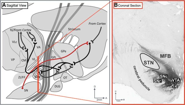
Figure 5.
Major anatomical pathways affected by STN stimulation. A, Diagram of the sagittal view of the location of the STN in relation to the medial forebrain bundle (red line) comprising the ascending dopaminergic nigrostriatal and mesocorticolimbic pathways. Diagram reprinted with permission from Devergnas and Wichmann, 2011. B, Representative coronal section of tyrosine hydroxylase immunohistochemical analysis of dopaminergic projections in relation to the STN (Gale et al., 2013). VTA, Ventral tegmental area.

