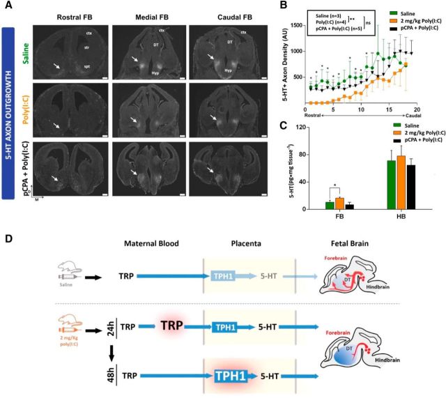
Figure 4.
Maternal inflammation disrupts fetal serotonergic axon outgrowth. A, IHC analysis of serotonergic axons in fetal brains 48 h after maternal exposure to either saline or 2 mg/kg poly(I:C) reveals blunted outgrowth of serotonergic axons in a caudal to rostral gradient within the fetal forebrain when comparing saline (control) with poly(I:C) treatments. This effect is not observed in the group coadministered with poly(I:C) and the TPH inhibitor pCPA [pCPA + poly(I:C); bottom panels]. B, Quantification of 5-HT+ axons density (normalized fluorescence intensity) throughout the rostrocaudal extent of E14 fetal forebrains obtained from saline (control)-, poly(I:C)-, or poly(I:C) + pCPA-treated dams 48 h after exposure. *p < 0.05, **p < 0.005, ns, not significant, Mann–Whitney U test. C, 5-HT is significantly increased in the fetal forebrain (FB) 48 h after maternal poly(I:C), but not poly(I:C) + pCPA injection; hindbrain (HB) 5-HT concentration is not affected significantlyin either group. *p < 0.05, unpaired t test with Holm–Sidak correction. n = 4 dams per group. D, Mild maternal inflammation induces a rapid increase in TRP metabolism through successively transient elevation of maternal blood/placental TRP concentration (24 h) followed by increased placental TPH1 enzymatic activity (48 h). This results in a sustained increase in fetal forebrain 5-HT tissue concentration and abnormal serotonergic axonal circuit formation. D, Dorsal; M, medial; Ctx, cortex; Str, striatum; Spt, septum; DT, dorsal thalamus; Hyp, hypothalamus. Error bars indicate SD. Scale bars, 200 μm.

