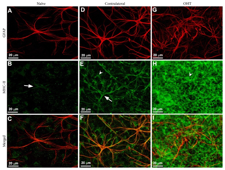
Figure 4.
Retinal macroglia in the mouse retina. Retinal whole-mount. Double immunostaining for GFAP (red) and MHC-II (green) after 15 days of laser-induced ocular hypertension. (A)–(C): naïve eyes; (D)–(F): contralateral eyes; (G)–(I): OHT eyes. In contralateral eyes, MHC-II immunoreaction of astrocytes (arrow) and Müller cells (arrowhead) in (E) was increased with respect to naïve eyes (arrow) in (B). In OHT eyes, MHC-II immunoreaction of Müller cells (arrowhead) in (H) was notably upregulated in comparison with contralateral (E). In OHT eyes the Müller cells were GFAP+ throughout the retina and appeared as punctate structures between the astrocytes and their radiating processes (G). Fluorescence microscopy and image acquisition using the ApoTome. GFAP: glial fibrillary acidic protein; MHC: major histocompatibility complex; OHT: ocular hypertension (from Figure 10 of [19] with permission).

