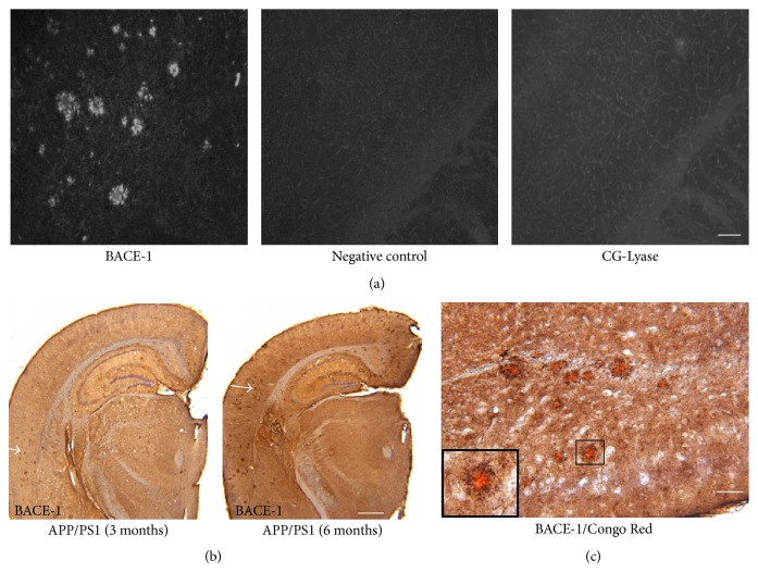
Figure 3.
BACE-1 protein accumulating in the brains of APP/PS1 mice. Representative photograph of immunofluorescent staining of BACE-1 protein in the brain cortex of APP/PS1 mice (a). BACE-1 accumulating in the brain of APP/PS1 mice at 3 and 6 months of age (b). Costaining with Congo Red and BACE-1 antibody in the brain cortex of APP/PS1 mice (c). Photograph in the black frame was magnified from the marked areas in the center. Scale bar represents 100 μm (a and c) and 500 μm (b).

