
Figure 9. Schematic diagram of the model .
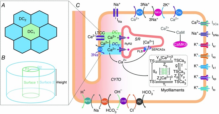
Human myocyte model (C) including ion pumps and exchangers responsible for action potential, Ca2+ management, force development, pHi and Ca2+–calmodulin‐dependent protein kinase II (CaMKII) regulation. Our previous model (Lascano et al. 2013) was modified by the addition of a second dyadic cleft: DC1 and DC2. The model is coupled to the myofilament force development model consisting of 5‐state troponin systems (TS) with Ca2+ binding at the myofilament level (Negroni et al. 2015). CYTO, cytosol; LTCC, L‐type Ca2+ channels; NCX, Na+/Ca2+ exchanger; NKA, Na+/K+‐ATPase; Cap, membrane Ca2+‐ATPase; NHE, Na+/H+ exchanger; NBC, Na+–HCO3 − cotransporter; CHE, Cl−/OH− exchanger; AE, anion exchanger; I represents different ionic currents (see text for further details). A, detail of the DC structure where DC1 is surrounded by 6 DC2. DC1 represents the dyadic clefts where RyR2 are facing the L‐type Ca2+ channel (LCC) and DC2 the dyadic clefts where RyR2 are not facing LCC. In the original TP model (ten Tusscher & Panfilov, 2006), the single dyadic cleft involves 14.3% of the present total cleft space (DC1+DC2). B, dyadic cleft structure assumed as two concentric cylinders. Surface 1: lateral surface of the cylinder with radius R, representing DC1. Surface 2: lateral surface of the cylinder with radius R2, representing DC2. The planar surfaces limiting the height of the cylinders are the sarcolemmal membrane on one side and the SR membrane on the other (see text for further details).