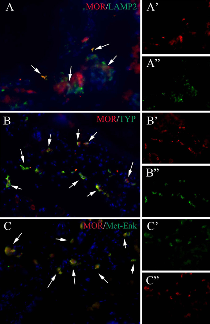
Fig. 3.
MOR is co-localized with MC marker, Lamp2, activated MC marker, TPSAB1, and with Met-Enk. a–a″ Typical image of MOR/Lamp2 double staining. a′, a″ Split channel of MOR and Lamp2, respectively. b–b″ Typical image of MOR/TPSAB1 double staining. b′, b″. Split channel of MOR and TPSAB1, respectively. c–c″ Typical image of MOR/Met-Enk double staining. c′, c″. Split channel of MOR and Met-Enk, respectively. Note that the co-localization of MOR and Lamp2 is only partial, whereas the co-localization of MOR/TPSAB1 and MOR/Met-Enk are almost exclusive

