
Figure 1:
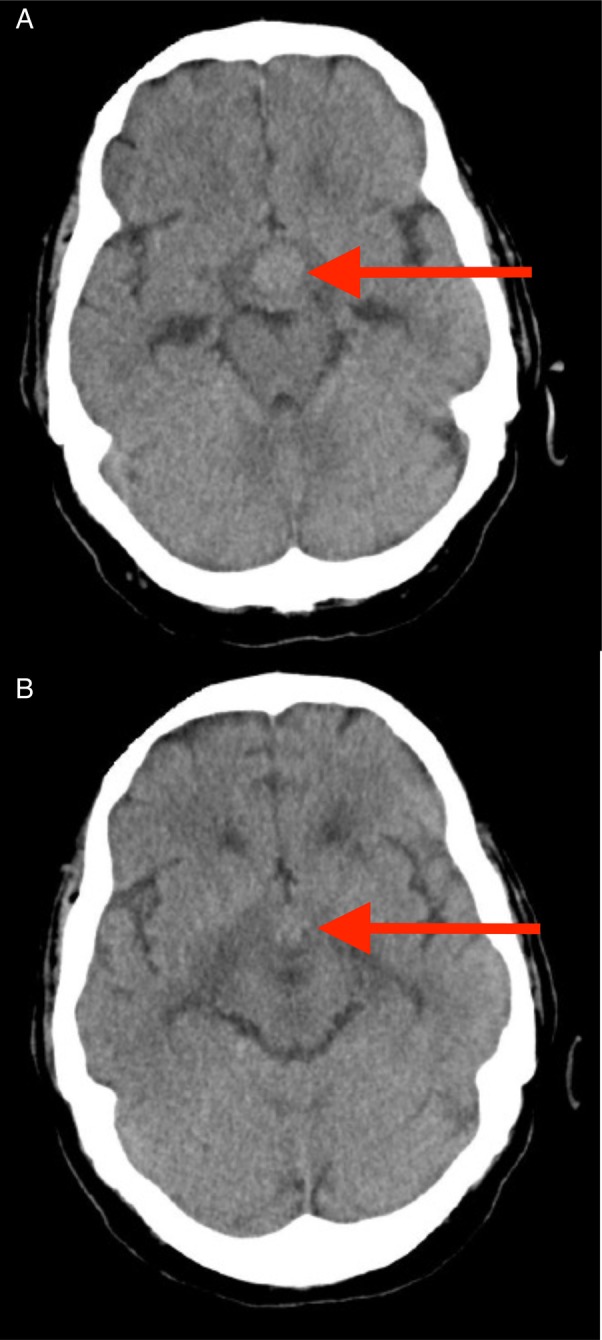
CT head scan images. An enhancing mass is seen in the pituitary fossa (Arrow, A). There is also overlying optic tract oedema (Arrow, B), not typically seen with pituitary adenomas.
Official websites use .gov
A
.gov website belongs to an official
government organization in the United States.
Secure .gov websites use HTTPS
A lock (
) or https:// means you've safely
connected to the .gov website. Share sensitive
information only on official, secure websites.
CT head scan images. An enhancing mass is seen in the pituitary fossa (Arrow, A). There is also overlying optic tract oedema (Arrow, B), not typically seen with pituitary adenomas.