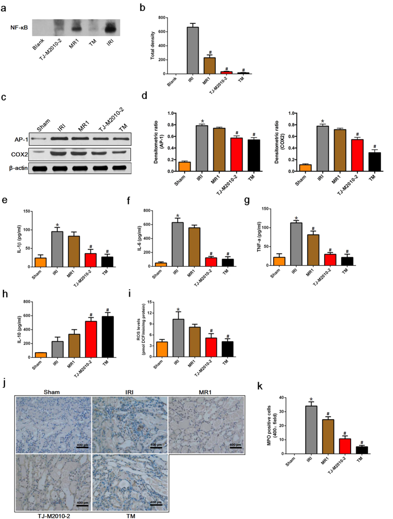
Figure 3. TJ-M2010-2 alone or with MR1 attenuates inflammatory responses after IRI.
Mice were exposed to IRI for 80 min with uninephrectomy. (a) Nuclear proteins were extracted from kidney tissues one day after renal IRI and incubated with an NF-κB probe for 25 min (three mice were sacrificed for each group). EMSA assay was used to detect NF-κB activity (one of three independent experiments). (b) Densitometric analysis of the NF-κB band in EMSA. (#p < 0.01 versus IRI). Results are expressed as mean ± s.d. Blank: no protein was added. (c) Total proteins were extracted from renal tissues one day after IRI. AP-1 and COX2 expression were measured by Western Blot. (one of three independent experiments). (d) Densitometric analysis of Western Blot results. The density of each β-actin lane was divided by that of AP-1 and COX2 (*p < 0.0001 versus Sham; #p < 0.0001 versus IRI). Results are expressed as mean ± s.d. (e–h) Serum samples were collected on day 1 after IRI (three of six mice sacrificed for the measurement of renal function for each group). IL1β, IL6, TNF-α and IL10 serum levels were quantified by ELISA. (*p < 0.01 versus Sham; #p < 0.01 versus IRI). Results are expressed as mean ± s.d. (i) Kidney tissues were obtained one day after IRI and homogenized to measure ROS formation. (*p < 0.01 versus Sham; #p < 0.01 versus IRI). Results are expressed as mean ± s.d. (j) Renal tissues were collected one day after IRI and stained by immunohistochemistry to detect MPO-positive cells (the same mice that were sacrificed for the assessment of pathologic damage were used for each group). Original magnification × 400 over five fields. MPO staining for neutrophil infiltration (brown: MPO-positive cells). Bar = 400 μm in all panels. (k) Semi-quantitative analysis of MPO positive cells. (*p < 0.01 versus Sham; #p < 0.01 versus IRI). Results are expressed as mean ± s.d.

