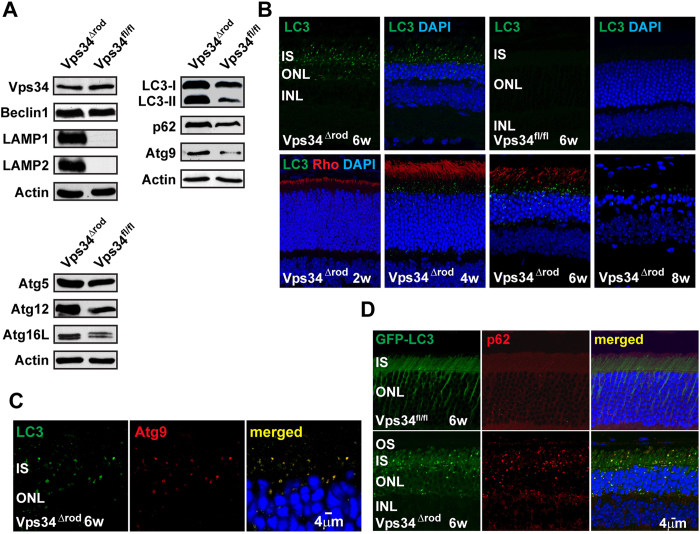
Figure 3. Dysfunction in the autophagy pathway in the absence of Vps34 function.
(A) Immunoblot reveals increased amounts of autophagy markers LC3/Atg8, Atg9, Atg12, Atg16L and p62, as well as lysosomal markers LAMP1 and LAMP2, in Vps34∆rod retinas. Both LC3-II levels and the ratio of LC3-II/LC3-I are increased. (B) LC3-staining puncta accumulated in rods at 4 weeks, prior to detectable changes in retinal structure. (C) LC3 co-localized with autophagosomal membrane marker Atg9 in inner segments of Vps34∆rod. (D) GFP-LC3 and p62 accumulated and co-localized in Vps34∆rod-LC3-GFP.

