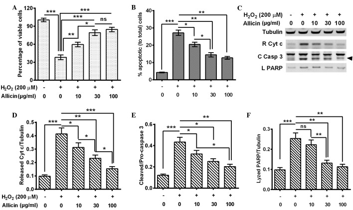
Figure 1.
Allicin inhibited H2O2-induced apoptosis in mouse osteoblastic MC3T3-E1 cells. MC3T3-E1 cells were treated with H2O2 (200 µM) for 6 h and 10, 30 or 100 µg/ml allicin for 24 h. (A) Cell viabilities. (B) Percentages of apoptotic cells; apoptotic cells were examined with an annexin V-FITC apoptosis detection kit. (C) Western blot analysis of cytochrome c release from mitochondria, cleaved caspase 3 (arrow) and lyzed PARP by caspase 3. (D) Ratio of released cytochrome c to tubulin. (E) Ratio of cleaved caspase 3 to pro-caspase 3. (F) Ratio of lyzed PARP to tubulin. All the experiments were performed in triplicate. *P<0.05, **P<0.01 or ***P<0.001, comparisons shown by brackets. ns, no significance; C Casp 3, cleaved caspase 3; L PARP, lyzed poly adenosine diphosphate-ribose polymerase; FITC, fluorescein isothiocyanate.

