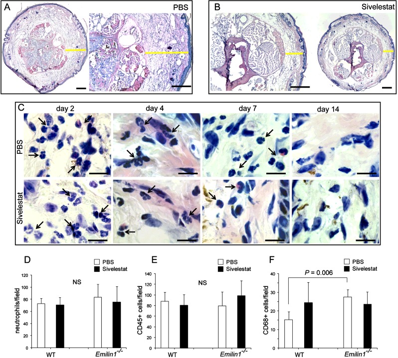
Figure 7. Sivelestat does not interfere with neutrophil recruitment.
(A, B) Yellow lines in representative cryostat sections after H&E staining indicate the dermis thickness of the tail 4 days after surgery in (A) control (PBS) and (B) sivelestat-treated mice. (C) High magnification images of the tail dermis show the presence of an abundant neutrophil infiltrate at days 2 and 4 in both control and treated mice, demonstrating that sivelestat administration does not inhibit the recruitment of neutrophils, which are also detectable up to day 7 after surgery. Black arrows indicate neutrophils. (D–F) Quantitative analyses of the number of (D) neutrophils, (E) CD45- and (F) CD68-positive cells in the tail 4 days after surgery in control (PBS) and sivelestat-treated WT and Emilin1−/− mice. The graphs report the means±S.D.s obtained from four mice per group, analysing eight fields (40×, original magnification) for each sample. The number of neutrophils was calculated on H&E-stained sections on the basis of morphological criteria, whereas the number of CD45- and CD68-positive cells was calculated on immunofluorescent sections. Scale bars=500 μm (A, B) and 20 μm (C).

