
Fig.1. Induction of DARPP-32 by RA does not require TrkB.
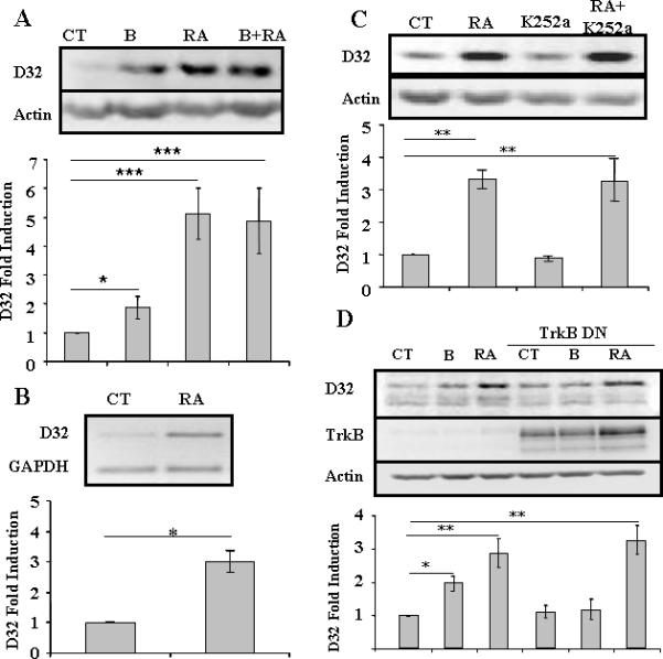
A) Primary striatal neurons were treated with BDNF (10 ng/ml) (B), retinoic acid (10 μM) (RA) or both for 24 hrs. Statistical analysis was performed using one-way ANOVA (p<0.0001) and significant differences were found between groups (*p<0.05 and ***p<0.001). (B) Primary striatal neurons were treated with RA (10 mM) for 24 hrs and levels of DARPP-32 and GAPDH mRNA were determined using semi-quantitative RT-PCR. Statistical analysis was performed using Student's T-test, * p< 0.01. (C) Primary striatal neurons were treated with RA (10 μM), K252a (100 nM) or both for 24 hrs. One-way ANOVA (p<0.001) and significant differences were found between groups (**p<0.01). (D) Primary striatal neurons were treated with RA (10 μM) in absence or presence of dominant negative TrkB (TrkB DN) adenovirus (m.o.i. = 2) for 24 hrs. Statistical analysis was performed using one-way ANOVA (p<0.0001) and significant differences were found between groups (*p<0.05 and **p<0.01) with the Bonferroni post-hoc test. (NOTE: TrkB was visible in wells without viral treatment upon over-exposure of the blot, data not shown).