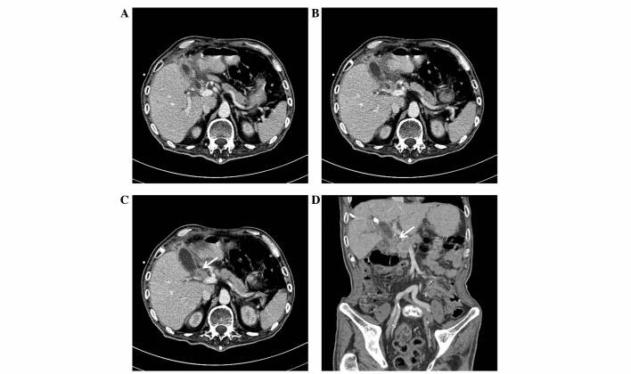
Figure 1.
Case 1. Contrast-enhanced computed tomography. (A) Mild dilation of the intrahepatic bile duct was observed, with a visible upper common bile duct and invisible middle and lower ducts. (B) Migration location between the dilated upper common bile duct and the tumor. (C) A clearly enhanced, higher density space-occupying lesion was present between the gastric antrum and duodenum (arrow). (D) Coronal reconstructed image showing the dilated common bile duct. The arrow indicates the lesion location.

