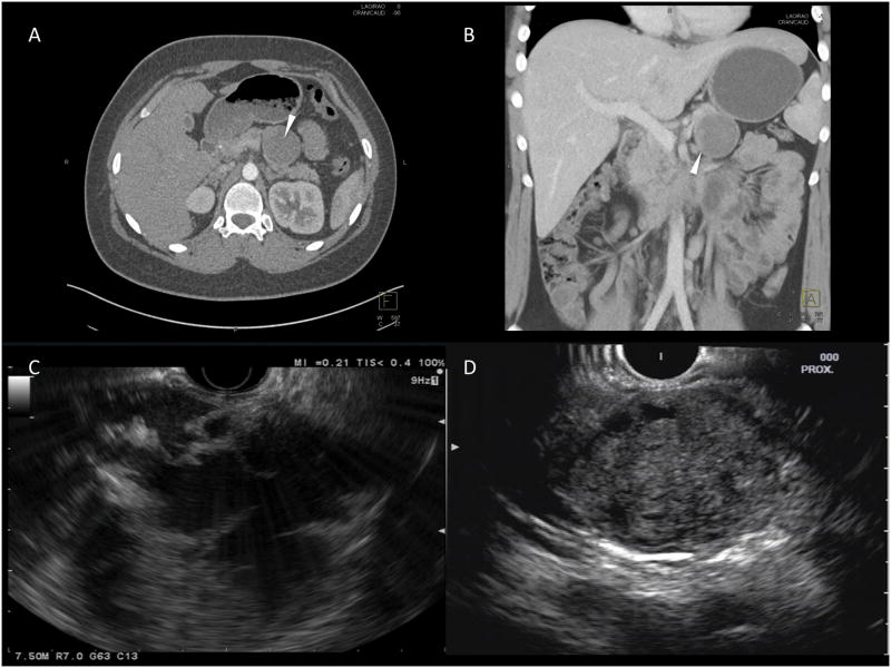
Figure 1. Imaging features of SPN.
This figure demonstrates the classic appearance of a SPN on imaging; (A) and (B) – CT images of mixed solid and cystic appearing lesion in the tail of the pancreas (arrowhead); (C) and (D) – EUS images demonstrating a mixed cystic-solid lesion (C) and a well-defined lesion with a mainly solid component (D).

