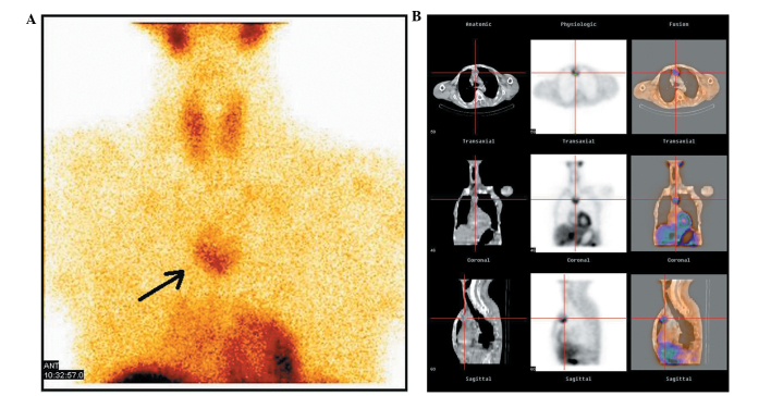
Figure 1.
Tc99m-MIBI scintigraphy scan of the patient's anterior mediastinum. (A) The static state of the parathyroid carcinoma. The black arrow indicates the tumor. (B) Three-dimensional reconstruction of parathyroid carcinoma. The center of the cross represents the location of the tumor. The first column corresponds to the anatomical scanning image by computed tomography. The second column is the physiological scan by Tc99m-MIBI scintigraphy. The third column represents the fusion of the first and second columns. The first line is the transaxial tomogram, the second line is the coronal tomogram and the third line corresponds to sagittal tomography. MIBI, methoxyisobutylisonitrile.

