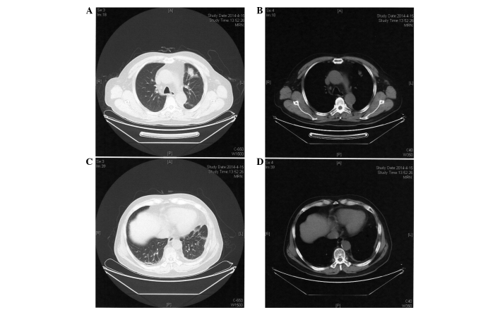
Figure 3.
Computed tomography scan performed on April 15, 2014 following 7 cycles of chemotherapy with pemetrexed and nedaplatin. (A) Lung and (B) mediastinal windows showing a mass in the upper lobe of the left lung and (C) lung and (D) mediastinal windows showing pleural effusions in the left side, which are decreased in size compared to those observed in Figure 1.

