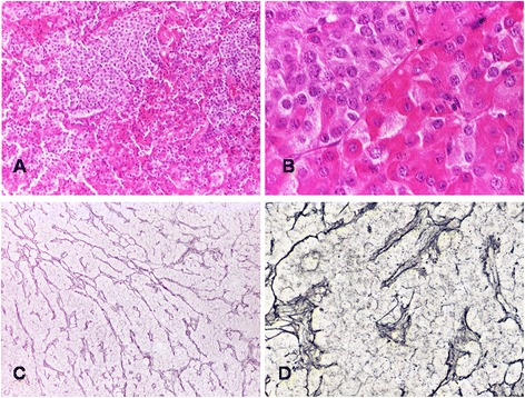
Fig. 2.

Adenomas occurring in XLAG patients are characterized by distinct populations of acidophilic and chromophobic cells (a HE - x10; b HE x40); staining for reticulin fibers highlights the lobular and cordonal architecture of XLAG-related adenomas (c Gordon-Sweet’s silver impregnation - x10); perivascular connective tissue containing thickened and distorted reticulin fibers (d Gordon-Sweet’s silver impregnation – x40)
