Fig. 3.

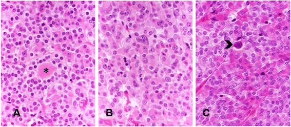
Secondary features of XLAG adenomas include pseudo-follicles containing colloid-like material (asterisk) (a HE – x20), isolated cells with large, irregular nucleus (b HE – x20), and scattered calcifications (arrow) (c HE – x20)

Secondary features of XLAG adenomas include pseudo-follicles containing colloid-like material (asterisk) (a HE – x20), isolated cells with large, irregular nucleus (b HE – x20), and scattered calcifications (arrow) (c HE – x20)