
Figure EV4. DDX6 represses translation initiation.
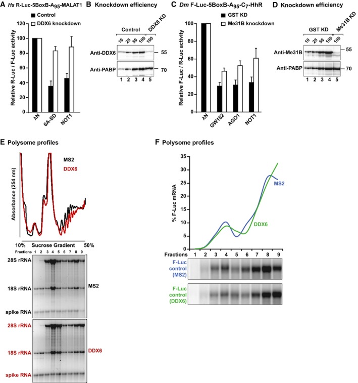
- Tethering assay using the R‐Luc‐5BoxB‐A95‐MALAT1 reporter and λN‐HA‐tagged TNRC6A‐SD and NOT1 in control HeLa cells or cells depleted of DDX6. Samples were analyzed as described in Fig 2. Error bars represent standard deviations from at least three independent experiments.
- Western blot showing DDX6 knockdown efficiency. Dilutions of control cell lysates were loaded in lanes 1–4 to estimate the efficacy of the depletion. PABP served as a loading control.
- Tethering assay using the F‐Luc‐5BoxB‐A95‐C7‐HhR reporter and λN‐HA‐tagged GW182, AGO1, and NOT1 in control S2 cells or cells depleted of Me31B. Samples were analyzed as described in Fig 2. Error bars represent standard deviations from at least three independent experiments.
- Western blot showing Me31B knockdown efficiency. Dilutions of control cell lysates were loaded in lanes 1–4 to estimate the efficacy of the depletion. PABP served as a loading control.
- Polysome profiles corresponding to Fig 7G. The absorbance at 254 nm of each fraction was quantitated and normalized to the total intensity across all fractions. The presence of 18S and 28S rRNAs in each fraction was analyzed on denaturing agarose gels. The spike RNA added to the fractions prior RNA isolation is indicated.
- The amount of F‐Luc control reporter corresponding to Fig 7G was quantified in each fraction of the gradient and normalized to the total amount across all fractions.
Source data are available online for this figure.