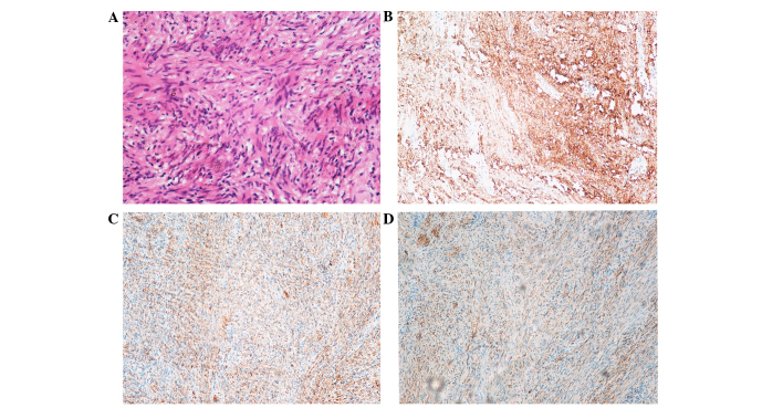
Figure 4.
(A) Hematoxylin and eosin staining revealed thin and long bipolar spindle cells with a typical palisading pattern. Hardly any mitotic figures were observed, and Antoni B areas characterized with less cellular, loosely textured Schwann cells were identified (original magnification, ×200). (B-D) Immumohistochemical staining showing positivity for (B) S100, (C) vimentin, (D) glial fibrillary acidic protein.

