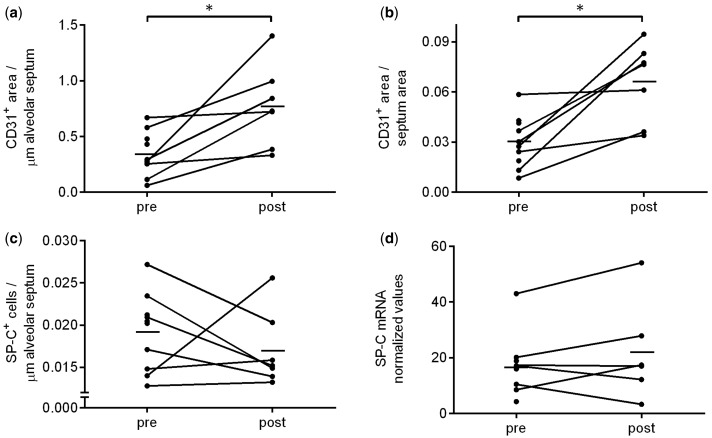
Figure 3.
CD31 and SP-C expression analysis in lung tissue before and after MSC infusion. Immunohistochemistry and mRNA expression performed on lung tissue obtained during first LVRS (pre) and a second LVRS procedure that was preceded by two MSC infusions (post). Quantification of CD31 IHC staining with (a) CD31 density, normalized by length of alveolar septa; and (b) CD31 area fraction, normalized by area of alveolar septa. (c) Quantification of IHC staining of SP-C (alveolar type II cell marker) normalized by length of alveolar septa. (d) mRNA expression of SP-C. Data in graph represent mean and individual data points. Paired data n = 7 for IHC and n = 6 for mRNA, * P < 0.05.

