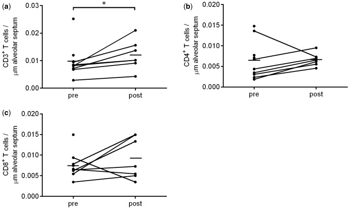
Figure 4.
IHC analysis of T cell markers pre- and post- MSC infusion. The number of CD3+, CD4+ and CD8+ T cells was assessed in surgical specimen obtained before and after MSC infusion. Quantification of (a) CD3, (b) CD4 and (c) CD8, expressed per length of alveolar septa. Data in graph represent mean (horizontal bar) and individual data points. Paired data n = 7, *P < 0.05.

