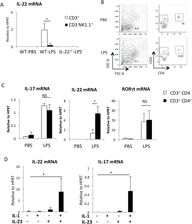
Fig. 6.
Detection of IL-22 in CD4+ T cells after LPS instillation in the lung and IL-22 production form memory CD4+ T cells in vitro. (A) Mononuclear cells isolated from the lungs 24h after PBS or LPS instillation were labeled with anti-CD3, anti-NK1.1 and anti-CD4 antibodies. Il22 mRNA expression in CD3+ T cells and NK cells (CD3-NK1.1+) FACS sorted from the lungs 24h after PBS or LPS instillation. n = 3 per group. *P < 0.05. (B) Representative data of flow cytometric analysis of cells isolated from the lungs 24h after PBS or LPS instillation. n > 5. (C) IL17, IL22 and RORγt mRNA expression in CD3+ CD4+ cells (Th cells) and CD3+ CD4- cells sorted from the lungs 24h after PBS or LPS instillation. mRNA levels were determined by quantitative RT–PCR. n = 3 per group. *P < 0.05. Values represent the mean ± SD. (D) Lung residential memory CD4+ T cells (CD4+CD44highCD62Llow) were purified by cell sorter and stimulated with IL-23 and/or IL-1β for 6h. mRNA levels of IL-22 and IL-17 were determined by quantitative RT–PCR.

