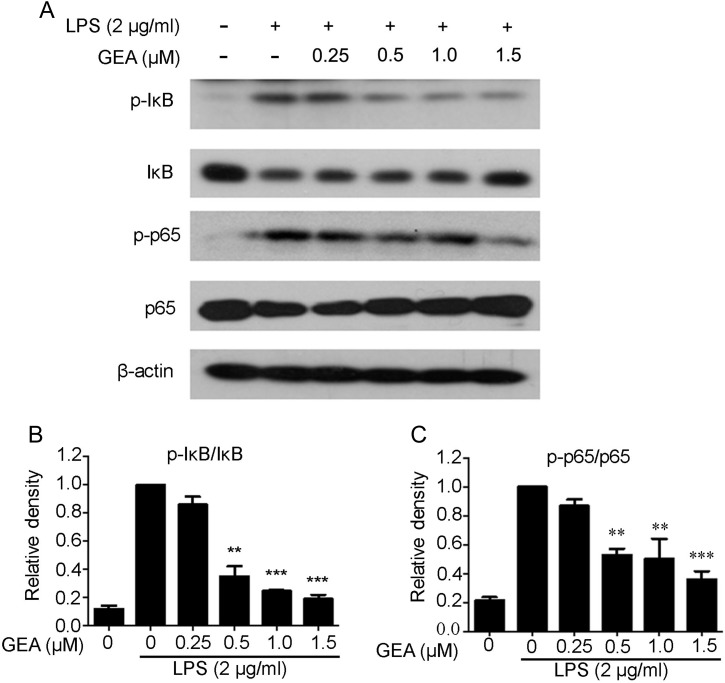
Figure 4.
Effect of GEA on LPS-induced NF-κB signaling in J774A.1 cells (A) Cells were pretreated with various concentrations of GEA for 1 h and then treated with 2 μg/ml LPS for another 1 h. The phosphorylation of p65 and IκBα was determined by western blot analysis using specific primary antibodies. (B) The band was quantified and expressed as the ratio of total p65 and IκBα intensities. Data are from three independent experiments. **P< 0.01 and ***P< 0.001 vs. LPS alone group.

