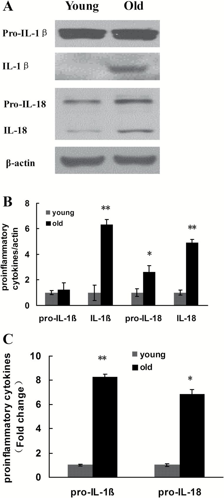
Figure 7.
Changes in the expression of pro-IL-1β, IL-1β, pro-IL-18, and IL-18 in the renal tissues of young and old rats. (A) Western blotting was used to detect the expression levels of proteins in each group of rats. (B) Quantitative gray scale analysis of protein levels. β-actin: internal reference. Compared with the young group, *indicates p < .05, **indicates p < .01. (C) Real-time–quantitative polymerase chain reaction detection results of pro-IL-1β and pro-IL-18. Compared with the young group, the fold changes in pro-IL-1β and pro-IL-18 in the old group = 2 − ΔΔCT. Compared with the young group, *indicates p < .05, **p < .01. IL-1βinterleukin-1β.

