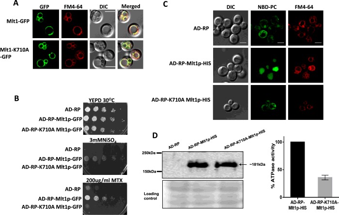
Figure 2. Expression, localization and characterization of WT Mlt1p and its mutant variant in a heterologous overexpressing strain.
(A) Fluorescence imaging by confocal microscope (right-hand panel) showing VM localization of Mlt1p–GFP and Mlt1p-K710A–GFP proteins with corresponding differential interference contrast (DIC) images, FM4-64 staining and merged images. Scale bar, 10 μm. (B) A comparison by spot dilution assays of susceptibilities of overexpressing strains AD-RP-Mlt1p-GFP, AD-RP-K710A Mlt1p-GFP with parental AD-RP strain. A 5-fold serial dilution of each strain was spotted on to NiSO4 and MTX at the indicated concentrations, in YEPD agar plates and grown for 48 h at 30°C. (C) NBD-PC accumulates in vacuolar lumen of WT Mlt1p-His-overexpressing strain (AD-RP-Mlt1p-HIS). Overexpression strains AD-RP-Mlt1p-HIS, AD-RP- K710A Mlt1p-HIS and parental strain AD-RP were grown to mid-exponential phase and incubated with NBD-PC. After incubation, cells were washed and stained with FM4-64. Samples were prepared for microscopy and photographed under a confocal microscope. Scale bar, 10 μm. (D) Immunoblot (left-hand panel) showing expression of Mlt1p-K710A–His and WT Mlt1p–His proteins. Vacuoles were isolated using the Ficoll gradient method and equal amounts of proteins (100 μg) were resolved by SDS/PAGE (8% gel) and then probed with anti-His antibody. After probing, membrane was stained with Ponceau S which is used as a loading control. ATPase activities (right-hand panel) in purified vacuolar vesicles from AD-RP-Mlt1p-HIS and AD-RP-Mlt1p-K710A-HIS strains were measured using an enzyme assay coupled to NADH oxidation at 340 nm. ATP hydrolysis is 60% reduced in Mlt1p-K710A-HIS mutants. Results are means±S.D. (n=3).

