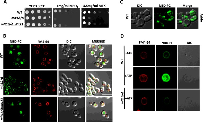
Figure 3. Mlt1p levels are required to maintain growth in the presence of NiSO4, methotrexate and for accumulation of NBD-PC into the vacuolar lumen.
(A) Comparison of growth by spot dilution assays of C. albicans WT (SC5314), mlt1Δ/Δ and mlt1Δ/Δ::MLT1 cells. A 5-fold serial dilution of each strain was spotted on to NiSO4 and MTX at the indicated concentrations, in YEPD agar plates and grown for 48 h at 30°C. (B) Deletion of MLT1 results in the loss of accumulation of NBD-PC in the vacuolar lumen. C. albicans WT (SC5314), mlt1Δ/Δ and mlt1Δ/Δ::MLT1 cells were grown to mid-exponential phase and incubated with NBD-PC. After incubation, cells were washed and stained with FM4-64. Samples were prepared for microscopy and photographed under a confocal microscope. Scale bar, 10 μm. (C) NBD-PC accumulation into the vacuolar lumen of C. albicans is energy-dependent. Sodium azide treatment was given to mid-exponential-phase WT C. albicans (SC5314) cells before their incubation with NBD-PC. The cells were photographed under a confocal microscope. (D) NBD-PC accumulation into the vacuolar lumen is energy-dependent. Vacuoles were isolated from WT and mlt1Δ/Δ mutant C. albicans using a Ficoll gradient ultracentrifuge-based method. Equal amounts (25 μg) of purified vacuolar vesicles were then incubated at 30°C for 30 min in buffer containing 10 μM NBD-PC and 10 μM FM4-64. A transport assay was carried out under two conditions: one in the absence of energy source ATP and another in the presence of 5 mM ATP. After incubation, samples were washed with ice-cold Tris/sucrose buffer containing 3% fatty-acid-free BSA and observed under a confocal microscope. In the presence of ATP, the isolated vacuoles from WT C. albicans showed NBD-PC accumulation. There was no NBD-PC accumulation in the absence of ATP in WT cells and in vacuoles isolated from mlt1Δ/Δ mutant. DIC, differential interference contrast.

