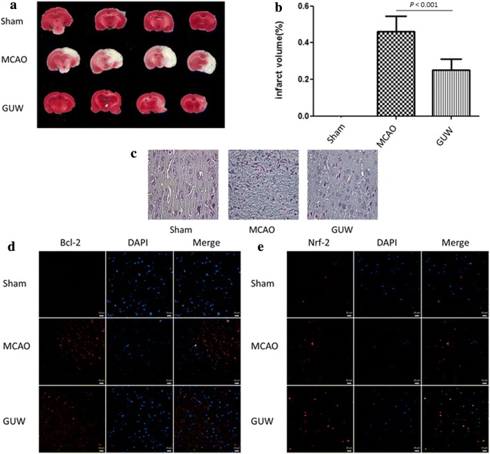
Fig. 6.

GUW reduced brain infarction volume and improved histological outcome. Brains were harvested 7 days of post-operation. The brains were sliced and performed TTC staining to evaluate the infarction volume and cresyl violet staining to examine the neuronal morphology at the infarct region. a The percentage of brain infarction of different groups of rats at 7 days of post-operation. b Data are expressed as mean ± SD, vs MCAO control group by one-way ANOVA (n = 10). c GUW increased the number of cells with normal neuronal morphology and decreased the number of shrunken and misshapen cells in cresyl violet–stained sections (magnification factor 400×). The Bcl-2 and Nrf-2 expression at the penumbra was examined by immunohistochemistry. GUW showed to upregulate both proteins in the penumbra. Representative images of the expression of d Bcl-2 and e Nrf-2. Scale bar 20 µm
