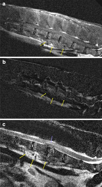
Fig. 1.

Magnetic resonance images of the lumbar spine of a male alpaca (Vicugna pacos). a T1-weighted sagittal MRI, b subtraction image and c T2-weighted sagittal MRI of the lumbar spine of a male alpaca. There is moderate enhancement of the endplates of L4/5, the ventral epidural space, and the sublumbar muscles. The endplates between the fourth and fifth lumbar vertebrae show irregular margins and the intervertebral disc space is narrowed. The intervertebral disc is degenerated and protrudes dorsally, leading to spinal cord compression. The myelon shows an irregular area of high signal intensity above the disc material
