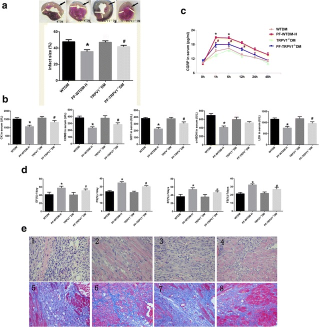
Fig. 2.

TRPV1 gene knockout partially abrogates paeoniflorin (PF)-mediated cardioprotection against MI. a TRPV1 knockout attenuated the decrease in myocardial infarct size by PF treatment, the black arrow indicates the infarction site. b TRPV1 knockout repressed the beneficial effects of PF on the release of myocardial enzymes in serum; c the activity of CGRP in serum measured at different time points before and after myocardial ischemia. d Heart function was assessed by fractional shortening {FS = [LV end-diastolic diameter (LVEDD) − LVend-systolic diameter (LVESD)] × 100/LVEDD} and ejection fraction [LVEF = (LVEDD2 − LVESD2)/LVEDD2]. e Representative images of hematoxylin and eosin (H&E, 1–4) and Masson’s trichrome staining (5–8); (1 and 5) WTDM group (2 and 6) PF-WTDM group, (3 and 7) TRPV1−/−DM group, (4 and 8) PF-TRPV1−/−DM. PF-WTDM group: WTDM mice were pretreated with PF (140 mg/kg) before surgery, and TRPV1−/−DM group: TRPV1 gene knockout mice with diabetes mellitus; PF-TRPV1−/−DM, TRPV1 gene knockout DM mice were pretreated with PF (140 mg/kg) before surgery; n = 6 per group; *P < 0.01, PF-TRPV1−/−WTDM vs. PF-WTDM-H; # P < 0.05, PF-TRPV1−/−DM vs. TRPV1−/−DM. (Mean ± SEM)
