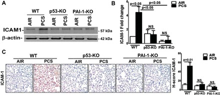
Fig. 4.

p53 and PAI-1 are prominently linked to PCS exposure-induced ICAM-1 expression. WT and p53- and PAI-1-deficient mice (n = 5/group) were kept in ambient air or exposed to PCS for 20 wk. A: lung homogenates prepared from these mice (n = 5) were immunoblotted for ICAM-1 expression. Representative image from triplicate analyses is showed. B: total RNA from the lung tissues of air or PCS mice (n = 5) were analyzed for ICAM-1 and β-actin mRNA. Bar graph represents mean ± SD of triplicate analyses. C: lung sections of air or PCS mice (n = 5) were subjected to IHC analysis for ICAM-1. Images are representative of IHC staining pattern of 10 fields (×200 magnification) and graph shows the H-scores.
