
Fig. 7.
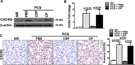
CSP inhibits PCS exposure induced pulmonary CXCR2 expression. WT mice were exposed to ambient air or PCS (n = 5/group) as described above. After 4 wk of PCS exposure, mice exposed to PCS were intraperitoneally injected with or without 18.75 mg/kg body wt of CSP or CP once every week for 4 more wk. After 20 wk of PCS exposure the mice were euthanized. A: lung homogenates (n = 5/group) were immunoblotted for CXCR2 and β-actin for loading equality. Representative image from triplicate analyses is showed. B: total RNA obtained from mice (n = 5/group) lung were analyzed for CXCR2 mRNA by RT-PCR as previously described. Bar graph represents mean ± SD of 3 independent analyses. C: lung sections from all groups of mice (n = 5/group) were subjected to IHC analysis using anti-CXCR2 antibody. Images are representative of IHC staining pattern of 10 fields (×200 magnification) and bar graph shows H-scores.