Fig. 6.

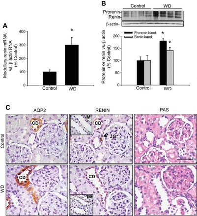
Renin expression is increased in the medullary collecting ducts following water deprivation. A: mice subjected to water deprivation (WD) showed increased renin mRNA levels in inner medullary tissues, which are not of juxtaglomerular origin. *P < 0.05 vs. control group; n = 4. B: prorenin and renin protein levels in inner medullary tissues were augmented by WD. *P < 0.05 vs. control group; n = 4. C: consecutive kidney sections (3 μm) from a control mouse (top) and a WD mouse (bottom) showing aquaporin-2 (AQP2), renin, and periodic acid-Schiff (PAS) staining. Kidney sections are from cortical regions to show juxtaglomerular (JG) renin as positive control (renin, top middle, arrow). Middle: renin staining in collecting ducts (CD) from cortex and inner medulla (IM; insets). Pictures were captured using a digital camera and an Eclipse Nikon microscope. Bar = 400 μm.
