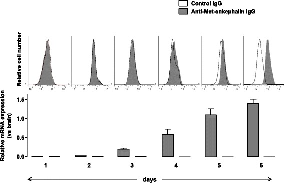
Fig. 2.

Proenkephalin is upregulated upon T cell activation. Naive CD4+ T lymphocytes isolated from wild-type mice were stimulated with both anti-CD3 and anti-CD28 mAbs for 6 days. Expression levels of mRNA encoding for PENK (gray histogram) and POMC (white histogram) were quantified by real-time PCR in CD4+ T lymphocytes from day 1 to day 6 (lower panels). mRNA content was normalized to the HPRT mRNA and quantified relative to standard mouse brain cDNA. Gene expression was assessed in at least five independent experiments run in duplicate. Results (mean ± SEM) are expressed relative to PENK or POMC mRNA expression in the mouse brain. Intracytoplasmic accumulation of Met-enkephalin-containing peptides was then assessed by cytofluorometry (upper panels). CD4+ T lymphocytes recovered each day of the stimulation were incubated with either control rabbit IgG (white histogram) or rabbit anti-Met-enkephalin IgG antibodies (gray histogram). The figure shows one representative experiment out of three performed
