Fig. 3.

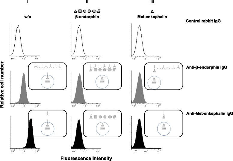
Anti-β-endorphin polyclonal IgG antibodies recognize enkephalins expressed in activated T lymphocytes as assessed by cytofluorometry. CD4+ T lymphocytes isolated from wild-type C57Bl/6 PENK+/+ mice were stimulated with both anti-CD3 and anti-CD28 mAbs for 6 days and intracellularly stained with control rabbit non-immune serum IgG (upper panels), rabbit anti-β-endorphin polyclonal IgG antibodies (middle panels), or rabbit anti-Met-enkephalin polyclonal IgG antibodies (lower panels). The figure depicts the binding of each of the three rabbit polyclonal IgG in the absence (i) or in the presence of an excess of soluble β-endorphin (ii) or Met-enkephalin (iii). The figure shows one representative experiment out of four performed. Schematic interpretation of the experiments is shown on the right of each histogram
